Mouse Exo-Check Exosomes Antibody Arrays
- Sample size: requires as little as 10µg of exosomal protein for detection (50 µg is optimal)
- Convenient: 8 known antibodies for exosome bona fide markers
- Flexibe: compatible with most exosome isolation methods, include the ExoQuick® family of reagents, SmartSEC, and ultracentrifugation
- Complete: includes a background control (blank spot) and a positive control for HRP validation
- Semi-quantitative: can be used to evaluate relative abundance of certain exosome markers from a given set of samples

Products
| Catalog Number | Description | Size | Price | Quantity | Add to Cart | |||
|---|---|---|---|---|---|---|---|---|
| EXORAY400A-4 | Mouse Exo-Check Exosome Antibody Array | 4 Arrays | $637 |
|
||||
| EXORAY400A-8 | Mouse Exo-Check Exosome Antibody Array | 8 Arrays | $1392 |
|
||||
Overview
Streamline your mouse exosome detection with Exo-Check Arrays For the most efficient detection of exosomes, turn to our semi-quantitative Exo-Check Exosome Antibody Arrays. Each array has 12 pre-printed spots and features 8 antibodies for known exosome markers (Cd63, Cd81, Alix, Flot1, Icam1, EpCam, Anxa5 and Tsg101), a Gm130 cis-Golgi matrix marker (Gm130), a labeled positive control for HRP detection, and a blank spot as a background control. The kits come complete with a secondary detection mixture conjugated to HRP.
- Sample size: requires as little as 10µg of exosomal protein for detection (50 µg is optimal)
- Convenient: 8 known antibodies for exosome bona fide markers
- Flexibe: compatible with most exosome isolation methods, include the ExoQuick® family of reagents, SmartSEC, and ultracentrifugation
- Complete: includes a background control (blank spot) and a positive control for HRP validation
- Semi-quantitative: can be used to evaluate relative abundance of certain exosome markers from a given set of samples
References
How It Works
A general reference to mouse Exo-Check Exosome Array
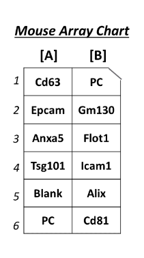
| Spot Position * | ID | NOTES | LINK TO EXOCARTA ENTRY FOR THE MOUSE PROTEINS |
|---|---|---|---|
| A1 | Cd63 | Tetraspanin | ExoCarta_12512 |
| A2 | Epcam | Epithelial call adhesion molecule; often found in cancer-derived exosomes | UniProt_Q99JW5 |
| A3 | Anxa5 | Annexin A5 | ExoCarta_11747 |
| A4 | Tsg101 | Tumor susceptibility gene 101 | ExoCarta_22088 |
| A5 | Blank | Background control | N/A |
| A6 | Positive Control | (+) control for HRP detection | N/A |
| B1 | Positive Control | (+) control for HRP detection | N/A |
| B2 | Gm130 | Cis-golgi matrix protein | N/A |
| B3 | Flot1 | Flotillin-1 | ExoCarta_14251 |
| B4 | Icam1 | Intercellular adhesion molecule 1 | ExoCarta_15894 |
| B5 | Alix | Programmed cell death 6 interacting protein (Pdcd6ip) | ExoCarta_18571 |
| B6 | Cd81 | Tetraspanin | ExoCarta_12520 |
Supporting Data
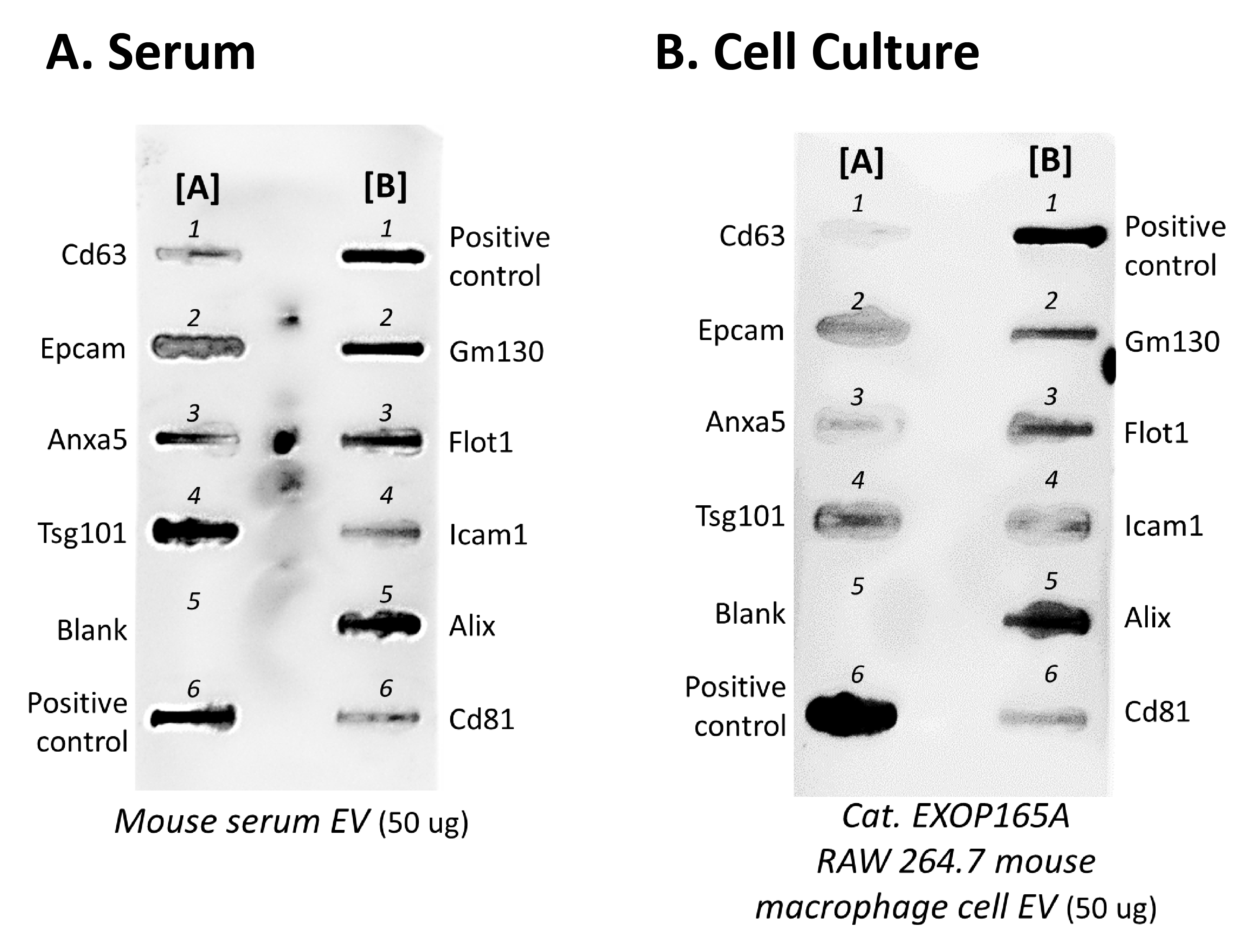
Figure 1. Serum- and cell culture-derived exosome analyzed with mouse Exo-Check Exosome Antibody Array
FAQs
Documentation
Citations
Related Products
Products
| Catalog Number | Description | Size | Price | Quantity | Add to Cart | |||
|---|---|---|---|---|---|---|---|---|
| EXORAY400A-4 | Mouse Exo-Check Exosome Antibody Array | 4 Arrays | $637 |
|
||||
| EXORAY400A-8 | Mouse Exo-Check Exosome Antibody Array | 8 Arrays | $1392 |
|
||||
Overview
Streamline your mouse exosome detection with Exo-Check Arrays For the most efficient detection of exosomes, turn to our semi-quantitative Exo-Check Exosome Antibody Arrays. Each array has 12 pre-printed spots and features 8 antibodies for known exosome markers (Cd63, Cd81, Alix, Flot1, Icam1, EpCam, Anxa5 and Tsg101), a Gm130 cis-Golgi matrix marker (Gm130), a labeled positive control for HRP detection, and a blank spot as a background control. The kits come complete with a secondary detection mixture conjugated to HRP.
- Sample size: requires as little as 10µg of exosomal protein for detection (50 µg is optimal)
- Convenient: 8 known antibodies for exosome bona fide markers
- Flexibe: compatible with most exosome isolation methods, include the ExoQuick® family of reagents, SmartSEC, and ultracentrifugation
- Complete: includes a background control (blank spot) and a positive control for HRP validation
- Semi-quantitative: can be used to evaluate relative abundance of certain exosome markers from a given set of samples
References
How It Works
A general reference to mouse Exo-Check Exosome Array
| Spot Position * | ID | NOTES | LINK TO EXOCARTA ENTRY FOR THE MOUSE PROTEINS |
|---|---|---|---|
| A1 | Cd63 | Tetraspanin | ExoCarta_12512 |
| A2 | Epcam | Epithelial call adhesion molecule; often found in cancer-derived exosomes | UniProt_Q99JW5 |
| A3 | Anxa5 | Annexin A5 | ExoCarta_11747 |
| A4 | Tsg101 | Tumor susceptibility gene 101 | ExoCarta_22088 |
| A5 | Blank | Background control | N/A |
| A6 | Positive Control | (+) control for HRP detection | N/A |
| B1 | Positive Control | (+) control for HRP detection | N/A |
| B2 | Gm130 | Cis-golgi matrix protein | N/A |
| B3 | Flot1 | Flotillin-1 | ExoCarta_14251 |
| B4 | Icam1 | Intercellular adhesion molecule 1 | ExoCarta_15894 |
| B5 | Alix | Programmed cell death 6 interacting protein (Pdcd6ip) | ExoCarta_18571 |
| B6 | Cd81 | Tetraspanin | ExoCarta_12520 |
Supporting Data
Figure 1. Serum- and cell culture-derived exosome analyzed with mouse Exo-Check Exosome Antibody Array