EXOCET Standards
- Fast—complete in 20 minutes
- Antibody-free—quantitation is based on the activity of AChE, an enzyme enriched in most exosomes
- Quantitative—included calibration standards enable calculation of the number of exosome particles
- Flexible—works with all mammalian species tested (human, mouse, rat)
- Fully compatible—works with most exosome isolation methods including the ExoQuick family of reagents, ultracentrifugation, immunoaffinity purification, and chromatography

Products
| Catalog Number | Description | Size | Price | Quantity | Add to Cart | |||
|---|---|---|---|---|---|---|---|---|
| EXOCET-SD-1 | EXOCET Standards | 1 Set | $267 |
|
||||
Overview
Overview
Extra standards for EXOCETWhile our fast and easy EXOCET Kit comes with standards, we also offer our EXOCET exosome standards as a stand-alone product for when you need more. The standards are calibrated using NanoSight analysis, enabling estimation of the number of exosomes in your samples.
The EXOCET Exosome Quantitation Kit is:
- Fast—complete in 20 minutes
- Antibody-free—quantitation is based on the activity of AChE, an enzyme enriched in most exosomes
- Quantitative—included calibration standards enable calculation of the number of exosome particles
- Flexible—works with all mammalian species tested (human, mouse, rat)
- Fully compatible—works with most exosome isolation methods including the ExoQuick family of reagents, ultracentrifugation, immunoaffinity purification, and chromatography
| ExoELISA-ULTRA Complete Kits | EXOCET | FluoroCet | |
|---|---|---|---|
| Use | For fast and sensitive antibody-based quantitation of exosomes | For fast quantitation of extracellular vesicles with moderate sample input requirements | For the most sensitive quantitation of extracellular vesicles with very low sample input requirements |
| Detection method | Antibody | Enzymatic | Enzymatic |
| Quantitation chemistry | Enzymatic (HRP) | Colorimetric | Fluorescent |
| Total protocol time | 4 hours (no overnight incubation) | 20 min | 60 min |
| Input sample amount (protein equivalent) | 1 – 200 µg | 50 µg | <1 µg |
| Learn More | ExoELISA-ULTRA CD63 ExoELISA-ULTRA CD81 ExoELISA-ULTRA CD9 ExoELISA-ULTRA GroEL | EXOCET | FluoroCet |
| ExoELISA-ULTRA CD63 Detection | ExoELISA CD9 ExoELISA CD63 ExoELISA CD81 | EXOCET | FluoroCet | |
| Use | For fast and sensitive antibody-based quantitation of exosomes | For sensitive quantitation of exosomes when time and input sample are not limiting | For fast quantitation of extracellular vesicles with moderate sample input requirements | For the most sensitive quantitation of extracellular vesicles with very low sample input requirements |
| Detection method | Antibody | Antibody | Enzymatic | Enzymatic |
| Quantitation chemistry | Enzymatic (HRP) | Enzymatic (HRP) | Colorimetric | Fluorescent |
| Total protocol time | 4 hours (no overnight incubation) | 24 hours | 20 min | 60 min |
| Input sample amount (protein equivalent) | 1 – 200 µg | >500 µg | 50 µg | <1 µg |
References
How It Works
How It Works
Exosome quantitation with EXOCET is quick and easy
- Isolate exosomes (ExoQuick, ExoQuick-TC, ExoQuick PLUS, and ExoQuick-TC PLUS are all excellent methods)
- Lyse exosomes with the included Exosome Lysis Buffer
- Measure AChE activity—add the included buffer and incubate for 10–20 minutes. Readout is at 405 nm
Supporting Data
Supporting Data
The fast, highly quantitative EXOCET exosome quantitation assay
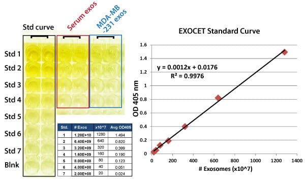
Exosome quantitation with EXOCET takes as little as 20 minutes and 50 µg (protein equivalent) of sample. Exosomes were isolated from either human serum (0.5 mL) or from MDA-MB-231 culture media (10 mL) using standard ExoQuick and ExoQuick-TC reagents. Exosome pellets were resuspended in PBS and total protein concentration measured using a BCA assay (~108 exosomes, 2 µg/µL). Exosomes were lysed using the EXOCET gentle lysis solution to preserve the enzymatic activity of the exosomal AChE enzyme. The standard curve was generated using known numbers of exosomes (as measured by NanoSight) and calibrated with a recombinant AChE enzyme standard solution provided in the kit.
FAQs
Documentation
Citations
Related Products
Products
| Catalog Number | Description | Size | Price | Quantity | Add to Cart | |||
|---|---|---|---|---|---|---|---|---|
| EXOCET-SD-1 | EXOCET Standards | 1 Set | $267 |
|
||||
Overview
Overview
Extra standards for EXOCETWhile our fast and easy EXOCET Kit comes with standards, we also offer our EXOCET exosome standards as a stand-alone product for when you need more. The standards are calibrated using NanoSight analysis, enabling estimation of the number of exosomes in your samples.
The EXOCET Exosome Quantitation Kit is:
- Fast—complete in 20 minutes
- Antibody-free—quantitation is based on the activity of AChE, an enzyme enriched in most exosomes
- Quantitative—included calibration standards enable calculation of the number of exosome particles
- Flexible—works with all mammalian species tested (human, mouse, rat)
- Fully compatible—works with most exosome isolation methods including the ExoQuick family of reagents, ultracentrifugation, immunoaffinity purification, and chromatography
| ExoELISA-ULTRA Complete Kits | EXOCET | FluoroCet | |
|---|---|---|---|
| Use | For fast and sensitive antibody-based quantitation of exosomes | For fast quantitation of extracellular vesicles with moderate sample input requirements | For the most sensitive quantitation of extracellular vesicles with very low sample input requirements |
| Detection method | Antibody | Enzymatic | Enzymatic |
| Quantitation chemistry | Enzymatic (HRP) | Colorimetric | Fluorescent |
| Total protocol time | 4 hours (no overnight incubation) | 20 min | 60 min |
| Input sample amount (protein equivalent) | 1 – 200 µg | 50 µg | <1 µg |
| Learn More | ExoELISA-ULTRA CD63 ExoELISA-ULTRA CD81 ExoELISA-ULTRA CD9 ExoELISA-ULTRA GroEL | EXOCET | FluoroCet |
| ExoELISA-ULTRA CD63 Detection | ExoELISA CD9 ExoELISA CD63 ExoELISA CD81 | EXOCET | FluoroCet | |
| Use | For fast and sensitive antibody-based quantitation of exosomes | For sensitive quantitation of exosomes when time and input sample are not limiting | For fast quantitation of extracellular vesicles with moderate sample input requirements | For the most sensitive quantitation of extracellular vesicles with very low sample input requirements |
| Detection method | Antibody | Antibody | Enzymatic | Enzymatic |
| Quantitation chemistry | Enzymatic (HRP) | Enzymatic (HRP) | Colorimetric | Fluorescent |
| Total protocol time | 4 hours (no overnight incubation) | 24 hours | 20 min | 60 min |
| Input sample amount (protein equivalent) | 1 – 200 µg | >500 µg | 50 µg | <1 µg |
References
How It Works
How It Works
Exosome quantitation with EXOCET is quick and easy
- Isolate exosomes (ExoQuick, ExoQuick-TC, ExoQuick PLUS, and ExoQuick-TC PLUS are all excellent methods)
- Lyse exosomes with the included Exosome Lysis Buffer
- Measure AChE activity—add the included buffer and incubate for 10–20 minutes. Readout is at 405 nm
Supporting Data
Supporting Data
The fast, highly quantitative EXOCET exosome quantitation assay
Exosome quantitation with EXOCET takes as little as 20 minutes and 50 µg (protein equivalent) of sample. Exosomes were isolated from either human serum (0.5 mL) or from MDA-MB-231 culture media (10 mL) using standard ExoQuick and ExoQuick-TC reagents. Exosome pellets were resuspended in PBS and total protein concentration measured using a BCA assay (~108 exosomes, 2 µg/µL). Exosomes were lysed using the EXOCET gentle lysis solution to preserve the enzymatic activity of the exosomal AChE enzyme. The standard curve was generated using known numbers of exosomes (as measured by NanoSight) and calibrated with a recombinant AChE enzyme standard solution provided in the kit.