Exosomes from Human Breast Milk, Normal (Single Donor)
- High purity and integrity
- Excellent reference for protein and RNA biomarkers
- Intact and fully functional
- Validated by NTA and Western Blot or ELISA

Products
| Catalog Number | Description | Size | Price | Quantity | Add to Cart | |||
|---|---|---|---|---|---|---|---|---|
| EXOP-540A-1 | Human Biofluids_Breast Milk, Healthy, Single Donor | 25 µg | $535 |
|
||||
Overview
Overview
Fuel your biomarker studies with purified exosomes from human biofluids. Whether you’re looking for exosome standards, performing functional studies, evaluating biomarkers, or engineering exosomes for therapeutic delivery, you can get your research moving faster with our ready-to-use exosomes isolated from the pooled serum of healthy human donors.- Ready to use—no validation required
- Lot-to-lot QC and validation data provided with every sales unit
- Fully standardized exosome controls
Our purified exosomes from pooled human biofluid samples come from healthy donors and include exosomes isolated from serum (Cat.# EXOP-500A-1), urine (Cat.# EXOP-520A-1), CSF (Cat.# EXOP-530A-1), and saliva (Cat.# EXOP-510A-1), with more biofluids on the way. Each lot of exosomes is carefully characterized for particle size and concentration by NanoSight™ analysis, and expression of specific exosome protein markers validated by Western blot.
- Protein biomarker analysis
- qPCR for RNA biomarkers
- High-throughput biomarker discovery (g. mass spec analysis)
- Electron microscopy
- Standardized controls for disease studies
Purified exosomes are stored in 1x PBS, and each unit contains 25 µg of exosomal protein. Lot-specific NanoSight and Western blot data are provided to ensure lot-to-lot quality and consistency. Each vial has enough material to run approximately 5 lanes in an SDS-PAGE gel (5 µg protein/lane).
Available purified exosomes| Cat.# | |
|---|---|
| Exosomes isolated from cancer cell lines | |
| EXOP-100A-1 | MCF-7 Human breast cancer, noninvasive cell line |
| EXOP-105A-1 | MDA-MB-231 Human breast cancer, aggressive/invasive/metastatic cell line |
| EXOP-115A-1 | PC-3 Human prostate cancer cells derived from metastatic cancer cell line |
| EXOP-120A-1 | A549 Human non-small cell lung cancer cell line |
| EXOP-125A-1 | H841 Human small cell lung cancer cell line |
| EXOP-130A-1 | H196 Human small cell lung cancer cell line |
| EXOP-135A-1 | DMS114 Human small cell lung cancer cell line |
| Exosomes isolated from stem cell lines | |
| EXOP-140A-1 | PCS-500-011 Human pre-adipose derived mesenchymal stem cell |
| EXOP-145A-1 | PCS-500-012 Human bone marrow-derived mesenchymal stem cell line |
| Exosomes isolated from immune-related cell lines | |
| EXOP-150A-1 | BC-3 Human B lymphocyte cell line |
| EXOP-155A-1 | Jurkat Clone E6-1 Human T lymphocyte cell line |
| EXOP-160A-1 | JM1 Human T pre-B lymphoblast cell line |
| EXOP-165A-1 | RAW 264.7 Mouse macrophage cell line |
| EXOP-200A-1 | JAWS II Mouse bone marrow immature dendritic cell line |
| Exosomes isolated from general cell lines | |
| EXOP-110A-1 | HEK293 Human embryonic kidney cell line |
| XPAK100EX-G | XPack-GFP-loaded HEK293 exosomes |
| Exosomes isolated from biofluid exosomes | |
| EXOP-500A-1 | Human pooled serum (healthy donors) |
| EXOP-510A-1 | Human pooled saliva (healthy donors) |
| EXOP-520A-1 | Human pooled urine (healthy donors) |
| EXOP-530A-1 | Human pooled CSF (healthy donors) |
| EXOP-540A-1 | Human breast milk, normal (single donor) |
| EXOP-550A-1 | Human biofluids ascites fluid (single donor) |
| Exosome isolated from mouse | |
| EXOP-560M-1 | Mouse Pooled Serum |
References
How It Works
Supporting Data
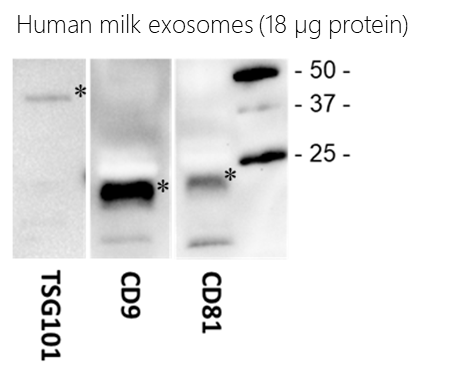
Figure 1. SBI’s Biofluid Exosomes contain expected protein markers as shown via Western blot analysis. The amount of protein loaded on each gel is as indicated.
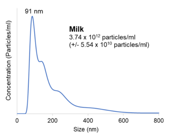
Figure 2. NanoSight analysis of Biofluid Exosomes show expected size distributions. Concentrations in particles/mL are shown, with the particle size mode reported on each plot.
FAQs
Documentation
Citations
Related Products
Products
| Catalog Number | Description | Size | Price | Quantity | Add to Cart | |||
|---|---|---|---|---|---|---|---|---|
| EXOP-540A-1 | Human Biofluids_Breast Milk, Healthy, Single Donor | 25 µg | $535 |
|
||||
Overview
Overview
Fuel your biomarker studies with purified exosomes from human biofluids. Whether you’re looking for exosome standards, performing functional studies, evaluating biomarkers, or engineering exosomes for therapeutic delivery, you can get your research moving faster with our ready-to-use exosomes isolated from the pooled serum of healthy human donors.- Ready to use—no validation required
- Lot-to-lot QC and validation data provided with every sales unit
- Fully standardized exosome controls
Our purified exosomes from pooled human biofluid samples come from healthy donors and include exosomes isolated from serum (Cat.# EXOP-500A-1), urine (Cat.# EXOP-520A-1), CSF (Cat.# EXOP-530A-1), and saliva (Cat.# EXOP-510A-1), with more biofluids on the way. Each lot of exosomes is carefully characterized for particle size and concentration by NanoSight™ analysis, and expression of specific exosome protein markers validated by Western blot.
- Protein biomarker analysis
- qPCR for RNA biomarkers
- High-throughput biomarker discovery (g. mass spec analysis)
- Electron microscopy
- Standardized controls for disease studies
Purified exosomes are stored in 1x PBS, and each unit contains 25 µg of exosomal protein. Lot-specific NanoSight and Western blot data are provided to ensure lot-to-lot quality and consistency. Each vial has enough material to run approximately 5 lanes in an SDS-PAGE gel (5 µg protein/lane).
Available purified exosomes| Cat.# | |
|---|---|
| Exosomes isolated from cancer cell lines | |
| EXOP-100A-1 | MCF-7 Human breast cancer, noninvasive cell line |
| EXOP-105A-1 | MDA-MB-231 Human breast cancer, aggressive/invasive/metastatic cell line |
| EXOP-115A-1 | PC-3 Human prostate cancer cells derived from metastatic cancer cell line |
| EXOP-120A-1 | A549 Human non-small cell lung cancer cell line |
| EXOP-125A-1 | H841 Human small cell lung cancer cell line |
| EXOP-130A-1 | H196 Human small cell lung cancer cell line |
| EXOP-135A-1 | DMS114 Human small cell lung cancer cell line |
| Exosomes isolated from stem cell lines | |
| EXOP-140A-1 | PCS-500-011 Human pre-adipose derived mesenchymal stem cell |
| EXOP-145A-1 | PCS-500-012 Human bone marrow-derived mesenchymal stem cell line |
| Exosomes isolated from immune-related cell lines | |
| EXOP-150A-1 | BC-3 Human B lymphocyte cell line |
| EXOP-155A-1 | Jurkat Clone E6-1 Human T lymphocyte cell line |
| EXOP-160A-1 | JM1 Human T pre-B lymphoblast cell line |
| EXOP-165A-1 | RAW 264.7 Mouse macrophage cell line |
| EXOP-200A-1 | JAWS II Mouse bone marrow immature dendritic cell line |
| Exosomes isolated from general cell lines | |
| EXOP-110A-1 | HEK293 Human embryonic kidney cell line |
| XPAK100EX-G | XPack-GFP-loaded HEK293 exosomes |
| Exosomes isolated from biofluid exosomes | |
| EXOP-500A-1 | Human pooled serum (healthy donors) |
| EXOP-510A-1 | Human pooled saliva (healthy donors) |
| EXOP-520A-1 | Human pooled urine (healthy donors) |
| EXOP-530A-1 | Human pooled CSF (healthy donors) |
| EXOP-540A-1 | Human breast milk, normal (single donor) |
| EXOP-550A-1 | Human biofluids ascites fluid (single donor) |
| Exosome isolated from mouse | |
| EXOP-560M-1 | Mouse Pooled Serum |
References
How It Works
Supporting Data
Figure 1. SBI’s Biofluid Exosomes contain expected protein markers as shown via Western blot analysis. The amount of protein loaded on each gel is as indicated.
Figure 2. NanoSight analysis of Biofluid Exosomes show expected size distributions. Concentrations in particles/mL are shown, with the particle size mode reported on each plot.