ExoGlow™-Membrane EV Labeling Kit
- Specific—carefully developed to generate a robust signal specific for EV membranes, leading to very low levels of background
- Compatible—delivers robust performance on EVs isolated using all methods tested—including ExoQuick®, ultracentrifugation, and column-based workflows
- Easy-to-use—labeling protocol is quick and straightforward
- Powerful—can be used with as little as 1 µg of EVs

Products
| Catalog Number | Description | Size | Price | Quantity | Add to Cart | |||
|---|---|---|---|---|---|---|---|---|
| EXOGM600A-1 | ExoGlowTM-Membrane EV Labeling Kit | 25 Reactions | $481 |
|
||||
Overview
Overview
See EVs better with reagents specifically optimized for labeling SBI’s new generation of extracellular vesicle (EV) labeling reagents take your EV visualization to new levels of clarity, with low background and high selectivity. Unlike general-purpose labeling reagents that are not optimized for EVs and suffer from high levels of background signal, ExoGlow™-Membrane improves your ability to track and localize EVs by specifically labeling EV membranes with a proprietary fluorescent dye that delivers very low levels of background signal. The result is unmatched EV imaging for more accurate studies (see below for our full range of next generation ExoGlow reagents).- Specific—carefully developed to generate a robust signal specific for EV membranes, leading to very low levels of background
- Compatible—delivers robust performance on EVs isolated using all methods tested—including ExoQuick®, ultracentrifugation, and column-based workflows
- Easy-to-use—labeling protocol is quick and straightforward
- Powerful—can be used with as little as 1 µg of EVs
Emission: 635 nm
Laser line: 488 nm Available next generation ExoGlow EV imaging reagents
| EV Component Labeled | Cat. # | Product | Excitation (nm) | Emission (nm) | Laser Line (nm) |
|---|---|---|---|---|---|
| Internal protein | EXOGP100A-1 | ExoGlow™-Protein EV Labeling Kit (Red) | 573 | 588 | 561 |
| EXOGP300A-1 | ExoGlow™-Protein EV Labeling Kit (Green) | 511 | 525 | 488 | |
| EXOGP400A-1 | ExoGlow™-Protein EV Labeling Kit (Blue) | 403 | 454 | 405 | |
| RNA | EXOGR800A-1 | ExoGlow™-RNA EV Labeling Kit | 485 | 537 | 488 |
| Membrane | EXOGM600A-1 | ExoGlow™-Membrane EV Labeling Kit | 465 | 635 | 488 |
References
How It Works
Supporting Data
Supporting Data
Imaging EVs with a new level of clarity
![]()
Figure 1. ExoGlow-Membrane enables clear visualization of labeled EVs being internalized by target cells. We labeled HEK293T EVs with ExoGlow-Membrane and followed uptake by HEK293T cells. The evenly distributed fluorescence signal shows internalization of labeled EVs by the target cells and the distribution of EV membranes to cellular membranes.
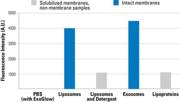
Figure 2. ExoGlow-Membrane is specific for intact membranes. We incubated either PBS, liposomes, liposomes pre-solubilized with detergent (0.05% Tx100), EVs, or lipoproteins. Only the samples with intact membranes—liposomes and EVs (blue)—show a high fluorescence signal, demonstrating ExoGlow-Membrane’s specificity for intact membranes.
FAQs
Documentation
Citations
Related Products
Products
| Catalog Number | Description | Size | Price | Quantity | Add to Cart | |||
|---|---|---|---|---|---|---|---|---|
| EXOGM600A-1 | ExoGlowTM-Membrane EV Labeling Kit | 25 Reactions | $481 |
|
||||
Overview
Overview
See EVs better with reagents specifically optimized for labeling SBI’s new generation of extracellular vesicle (EV) labeling reagents take your EV visualization to new levels of clarity, with low background and high selectivity. Unlike general-purpose labeling reagents that are not optimized for EVs and suffer from high levels of background signal, ExoGlow™-Membrane improves your ability to track and localize EVs by specifically labeling EV membranes with a proprietary fluorescent dye that delivers very low levels of background signal. The result is unmatched EV imaging for more accurate studies (see below for our full range of next generation ExoGlow reagents).- Specific—carefully developed to generate a robust signal specific for EV membranes, leading to very low levels of background
- Compatible—delivers robust performance on EVs isolated using all methods tested—including ExoQuick®, ultracentrifugation, and column-based workflows
- Easy-to-use—labeling protocol is quick and straightforward
- Powerful—can be used with as little as 1 µg of EVs
Emission: 635 nm
Laser line: 488 nm Available next generation ExoGlow EV imaging reagents
| EV Component Labeled | Cat. # | Product | Excitation (nm) | Emission (nm) | Laser Line (nm) |
|---|---|---|---|---|---|
| Internal protein | EXOGP100A-1 | ExoGlow™-Protein EV Labeling Kit (Red) | 573 | 588 | 561 |
| EXOGP300A-1 | ExoGlow™-Protein EV Labeling Kit (Green) | 511 | 525 | 488 | |
| EXOGP400A-1 | ExoGlow™-Protein EV Labeling Kit (Blue) | 403 | 454 | 405 | |
| RNA | EXOGR800A-1 | ExoGlow™-RNA EV Labeling Kit | 485 | 537 | 488 |
| Membrane | EXOGM600A-1 | ExoGlow™-Membrane EV Labeling Kit | 465 | 635 | 488 |
References
How It Works
Supporting Data
Supporting Data
Imaging EVs with a new level of clarity
![]()
Figure 1. ExoGlow-Membrane enables clear visualization of labeled EVs being internalized by target cells. We labeled HEK293T EVs with ExoGlow-Membrane and followed uptake by HEK293T cells. The evenly distributed fluorescence signal shows internalization of labeled EVs by the target cells and the distribution of EV membranes to cellular membranes.
Figure 2. ExoGlow-Membrane is specific for intact membranes. We incubated either PBS, liposomes, liposomes pre-solubilized with detergent (0.05% Tx100), EVs, or lipoproteins. Only the samples with intact membranes—liposomes and EVs (blue)—show a high fluorescence signal, demonstrating ExoGlow-Membrane’s specificity for intact membranes.