Exo-FITC Exosome FACS Stain

Products
| Catalog Number | Description | Size | Price | Quantity | Add to Cart | |||
|---|---|---|---|---|---|---|---|---|
| EXOFLOW800A-1 | Exo-FITC Exosome FACS stain | 20 Reactions | $195 |
|
||||
Overview
Overview
Visualize bead-immobilized exosomes with our proprietary, reversible FITC-based stain
Optimized for our Exo-Flow Capture Kits and available only from SBI, Exo-FITC is a proprietary reversible fluorescent stain for visualizing exosomes. Exo-FITC is comprised of the commonly used fluorophore fluorescein isothiocyanate (FITC) conjugated to a protein that recognizes post-translational modifications on exosomal surface proteins, enabling general staining of exosomes.
However, the feature that really makes Exo-FITC stand out as an ideal reagent for FACS-based isolation of exosomes is its reversibility—after staining with Exo-FITC and flow-sorting by FITC-positive gating (Exo-FITC’s excitation and emission wavelengths are 494 nm and 518 nm, respectively), Exo-FITC can be removed from exosomes through the simple addition of Exosome Elution Buffer, which simultaneously elutes exosomes from the Exo-Flow magnetic beads and removes bound Exo-FITC.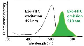
Note that we also offer reversible Exo-APC (Cat.# Exo-FLOW810A-1) as an alternative stain to Exo-FITC.
Use Exo-FITC with any of our Exo-Flow Capture Kits:| Cat.# | Kit |
|---|---|
| EXOFLOW100A-1 | CD9 Exo-Flow Capture Kit |
| EXOFLOW150A-1 | Tetraspanin Exo-Flow Combo Capture Kit |
| EXOFLOW200A-1 | CD31 Exo-Flow Capture Kit |
| EXOFLOW210A-1 | CD44 Exo-Flow Capture Kit |
| EXOFLOW300A-1 | CD63 Exo-Flow Capture Kit |
| EXOFLOW400A-1 | CD81 Exo-Flow Capture Kit |
| EXOFLOW500A-1 | Rab5b Exo-Flow Capture Kit |
| EXOFLOW600A-1 | HLA-G Exo-Flow Capture Kit |
| EXOFLOW610A-1 | CD14 Exo-Flow Capture Kit |
| EXOFLOW620A-1 | CD68 Exo-Flow Capture Kit |
| EXOFLOW630A-1 | EpCAM Exo-Flow Capture Kit |
| EXOFLOW660A-1 | CD73 Exo-Flow Capture Kit |
How It Works
How It Works
Easily purify exosomes using FACS with our Exo-Flow Capture Kits
At a glance
Simply (1) couple biotinylated antibody to the magnetic streptavidin beads, (2) use the antibody-coupled magnetic beads to capture exosomes that have been isolated using either ExoQuick® or ultracentrifugation, (3) wash away unbound exosomes, and then (4) stain with reversible Exo-FITC (excitation and emission wavelengths of 494 nm and 518 nm, respectively).
Your sample is now ready for FACS analysis.
To use the purified exosomes after FACS, add the included Exosome Elution Buffer to simultaneously remove the Exo-FITC stain and elute intact exosomes from the beads.
Supporting Data
Supporting Data
See Exo-FITC in action
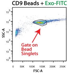
Exo-FITC enables selective exosome isolation via FACS. Exosomes are isolated using the CD9 Exo-Flow Capture Kit and flow-sorted using FITC-positive gating.
FAQs
Resources
Related Products
Citations
-
Boyer, E, et al. (2024) Comparison of plasma soluble and extracellular vesicles-associated biomarkers in Alzheimer’s Disease patients and cognitively normal individuals. medRxiv. 2024;. Link: medRxiv
-
Klemetti, MM, et al. (2024) Lipid profile of circulating placental extracellular vesicles during pregnancy identifies foetal growth restriction risk. Journal of extracellular vesicles. 2024; 13(2):e12413. PM ID: 38353485
-
Mekala, N, et al. (2024) Alcohol and e-cigarette damage alveolar-epithelial barrier by activation of P2X7r and provoke brain endothelial injury via extracellular vesicles. Cell communication and signaling : CCS. 2024; 22(1):39. PM ID: 38225580
-
Garbin, A, et al. (2023) MiR-146a-5p enrichment in small-extracellular vesicles of relapsed pediatric ALCL patients promotes macrophages infiltration and differentiation. Biochemical pharmacology. 2023; 215:115747. PM ID: 37591448
-
Guda, P, et al. (2023) Nanoscopic and functional characterization of keratinocyte-originating exosomes in the wound fluid of non-diabetic and diabetic chronic wound patients. Nano Today. 2023; 52:101954. Link: Nano Today
-
Nakazaki, M, et al. (2023) Human mesenchymal stem-derived extracellular vesicles improve body growth and motor function following severe spinal cord injury in rat. Clinical and translational medicine. 2023; 13(6):e1284. PM ID: 37323108
-
Mimmi, S, et al. (2023) SARS CoV-2 spike protein-guided exosome isolation facilitates detection of potential miRNA biomarkers in COVID-19 infections. Clinical chemistry and laboratory medicine. 2023;. PM ID: 36972680
-
Bertolini, I, et al. (2023) Intercellular HIF1a reprogams mammary progenitors and myeloid immune evasion to drive high-risk breast lesions. The Journal of clinical investigation. 2023;. PM ID: 36892943
-
Shen, H & Lane, RA. (2023) Extracellular Vesicles from Inflammation-Primed Adipose-Derived Stem Cells Enhance Achilles Tendon Repair by Reducing Inflammation and Promoting Intrinsic Healing. bioRxiv : the preprint server for biology. 2023;. PM ID: 36778262
-
Yuan, X, et al. (2023) Contribution of Hepatic Steatosis-Intensified Extracellular Vesicle Release to Aggravated Inflammatory Endothelial Injury in Liver-Specific Asah1 Gene Knockout Mice. The American journal of pathology. 2023;. PM ID: 36638912
-
Koken, GY, et al. (2022) Wharton Jelly Derived Mesenchymal Stem Cell’s Exosomes Demonstrate Significant Antileishmanial and Wound Healing Effects in Combination with Aloe-Emodin: An in Vitro Study. Journal of pharmaceutical sciences. 2022;. PM ID: 35995206
-
Luo, Z, et al. (2022) Human bone marrow mesenchymal stem cell-derived extracellular vesicles inhibit shoulder stiffness via let-7a/Tgfbr1 axis. Bioactive Materials. 2022;. Link: Bioactive Materials
-
Nirujogi, TS, et al. (2022) Lipidomic Profiling of Bronchoalveolar Lavage Fluid Extracellular Vesicles Indicates Their Involvement in Lipopolysaccharide-Induced Acute Lung Injury. Journal of innate immunity. 2022;:1-14. PM ID: 35367992
-
Marra, KV, et al. (2022) Bioactive extracellular vesicles from a subset of endothelial progenitor cells rescue retinal ischemia and neurodegeneration. JCI insight. 2022; 7(12). PM ID: 35639473
-
Li, Y, et al. (2022) Exosome-shuttled miR-126 mediates ethanol-induced disruption of neural crest cell-placode cell interaction by targeting SDF1. Research Square. 2022;. Link: Research Square
-
Mullen, M, et al. (2022) Mechanical strain drives exosome production, function, and miRNA cargo in C2C12 muscle progenitor cells. Journal of orthopaedic research : official publication of the Orthopaedic Research Society. 2022;. PM ID: 36250617
-
Beatriz, M, et al. (2022) Defective mitochondria‐lysosomal axis enhances the release of extracellular vesicles containing mitochondrial DNA and proteins in Huntington’s disease. Journal of Extracellular Biology. 2022; 1(10). Link: Journal of Extracellular Biology
-
Zhang, H, et al. (2021) Improving Isolation of Extracellular Vesicles by Utilizing Nanomaterials. Membranes. 2021; 12(1). PM ID: 35054584
-
Luo, ZW, et al. (2021) Exosomes derived from inflammatory myoblasts promote M1 polarization and break the balance of myoblast proliferation/differentiation. World journal of stem cells. 2021; 13(11):1762-1782. PM ID: 34909122
-
Zhang, Z, et al. (2021) Surface Located Adiponectin On Adipocytes-derived Exosomes Mediates Adipocytes/cardiomyocytes Communication And Contributes To Cardioprotection. Circulation. 2021;. Link: Circulation
- See More
Products
| Catalog Number | Description | Size | Price | Quantity | Add to Cart | |||
|---|---|---|---|---|---|---|---|---|
| EXOFLOW800A-1 | Exo-FITC Exosome FACS stain | 20 Reactions | $195 |
|
||||
Overview
Overview
Visualize bead-immobilized exosomes with our proprietary, reversible FITC-based stain
Optimized for our Exo-Flow Capture Kits and available only from SBI, Exo-FITC is a proprietary reversible fluorescent stain for visualizing exosomes. Exo-FITC is comprised of the commonly used fluorophore fluorescein isothiocyanate (FITC) conjugated to a protein that recognizes post-translational modifications on exosomal surface proteins, enabling general staining of exosomes.
However, the feature that really makes Exo-FITC stand out as an ideal reagent for FACS-based isolation of exosomes is its reversibility—after staining with Exo-FITC and flow-sorting by FITC-positive gating (Exo-FITC’s excitation and emission wavelengths are 494 nm and 518 nm, respectively), Exo-FITC can be removed from exosomes through the simple addition of Exosome Elution Buffer, which simultaneously elutes exosomes from the Exo-Flow magnetic beads and removes bound Exo-FITC.
Note that we also offer reversible Exo-APC (Cat.# Exo-FLOW810A-1) as an alternative stain to Exo-FITC.
Use Exo-FITC with any of our Exo-Flow Capture Kits:| Cat.# | Kit |
|---|---|
| EXOFLOW100A-1 | CD9 Exo-Flow Capture Kit |
| EXOFLOW150A-1 | Tetraspanin Exo-Flow Combo Capture Kit |
| EXOFLOW200A-1 | CD31 Exo-Flow Capture Kit |
| EXOFLOW210A-1 | CD44 Exo-Flow Capture Kit |
| EXOFLOW300A-1 | CD63 Exo-Flow Capture Kit |
| EXOFLOW400A-1 | CD81 Exo-Flow Capture Kit |
| EXOFLOW500A-1 | Rab5b Exo-Flow Capture Kit |
| EXOFLOW600A-1 | HLA-G Exo-Flow Capture Kit |
| EXOFLOW610A-1 | CD14 Exo-Flow Capture Kit |
| EXOFLOW620A-1 | CD68 Exo-Flow Capture Kit |
| EXOFLOW630A-1 | EpCAM Exo-Flow Capture Kit |
| EXOFLOW660A-1 | CD73 Exo-Flow Capture Kit |
How It Works
How It Works
Easily purify exosomes using FACS with our Exo-Flow Capture Kits
At a glance
Simply (1) couple biotinylated antibody to the magnetic streptavidin beads, (2) use the antibody-coupled magnetic beads to capture exosomes that have been isolated using either ExoQuick® or ultracentrifugation, (3) wash away unbound exosomes, and then (4) stain with reversible Exo-FITC (excitation and emission wavelengths of 494 nm and 518 nm, respectively).
Your sample is now ready for FACS analysis.
To use the purified exosomes after FACS, add the included Exosome Elution Buffer to simultaneously remove the Exo-FITC stain and elute intact exosomes from the beads.
Supporting Data
Supporting Data
See Exo-FITC in action
Exo-FITC enables selective exosome isolation via FACS. Exosomes are isolated using the CD9 Exo-Flow Capture Kit and flow-sorted using FITC-positive gating.
FAQs
Citations
-
Boyer, E, et al. (2024) Comparison of plasma soluble and extracellular vesicles-associated biomarkers in Alzheimer’s Disease patients and cognitively normal individuals. medRxiv. 2024;. Link: medRxiv
-
Klemetti, MM, et al. (2024) Lipid profile of circulating placental extracellular vesicles during pregnancy identifies foetal growth restriction risk. Journal of extracellular vesicles. 2024; 13(2):e12413. PM ID: 38353485
-
Mekala, N, et al. (2024) Alcohol and e-cigarette damage alveolar-epithelial barrier by activation of P2X7r and provoke brain endothelial injury via extracellular vesicles. Cell communication and signaling : CCS. 2024; 22(1):39. PM ID: 38225580
-
Garbin, A, et al. (2023) MiR-146a-5p enrichment in small-extracellular vesicles of relapsed pediatric ALCL patients promotes macrophages infiltration and differentiation. Biochemical pharmacology. 2023; 215:115747. PM ID: 37591448
-
Guda, P, et al. (2023) Nanoscopic and functional characterization of keratinocyte-originating exosomes in the wound fluid of non-diabetic and diabetic chronic wound patients. Nano Today. 2023; 52:101954. Link: Nano Today
-
Nakazaki, M, et al. (2023) Human mesenchymal stem-derived extracellular vesicles improve body growth and motor function following severe spinal cord injury in rat. Clinical and translational medicine. 2023; 13(6):e1284. PM ID: 37323108
-
Mimmi, S, et al. (2023) SARS CoV-2 spike protein-guided exosome isolation facilitates detection of potential miRNA biomarkers in COVID-19 infections. Clinical chemistry and laboratory medicine. 2023;. PM ID: 36972680
-
Bertolini, I, et al. (2023) Intercellular HIF1a reprogams mammary progenitors and myeloid immune evasion to drive high-risk breast lesions. The Journal of clinical investigation. 2023;. PM ID: 36892943
-
Shen, H & Lane, RA. (2023) Extracellular Vesicles from Inflammation-Primed Adipose-Derived Stem Cells Enhance Achilles Tendon Repair by Reducing Inflammation and Promoting Intrinsic Healing. bioRxiv : the preprint server for biology. 2023;. PM ID: 36778262
-
Yuan, X, et al. (2023) Contribution of Hepatic Steatosis-Intensified Extracellular Vesicle Release to Aggravated Inflammatory Endothelial Injury in Liver-Specific Asah1 Gene Knockout Mice. The American journal of pathology. 2023;. PM ID: 36638912
-
Koken, GY, et al. (2022) Wharton Jelly Derived Mesenchymal Stem Cell’s Exosomes Demonstrate Significant Antileishmanial and Wound Healing Effects in Combination with Aloe-Emodin: An in Vitro Study. Journal of pharmaceutical sciences. 2022;. PM ID: 35995206
-
Luo, Z, et al. (2022) Human bone marrow mesenchymal stem cell-derived extracellular vesicles inhibit shoulder stiffness via let-7a/Tgfbr1 axis. Bioactive Materials. 2022;. Link: Bioactive Materials
-
Nirujogi, TS, et al. (2022) Lipidomic Profiling of Bronchoalveolar Lavage Fluid Extracellular Vesicles Indicates Their Involvement in Lipopolysaccharide-Induced Acute Lung Injury. Journal of innate immunity. 2022;:1-14. PM ID: 35367992
-
Marra, KV, et al. (2022) Bioactive extracellular vesicles from a subset of endothelial progenitor cells rescue retinal ischemia and neurodegeneration. JCI insight. 2022; 7(12). PM ID: 35639473
-
Li, Y, et al. (2022) Exosome-shuttled miR-126 mediates ethanol-induced disruption of neural crest cell-placode cell interaction by targeting SDF1. Research Square. 2022;. Link: Research Square
-
Mullen, M, et al. (2022) Mechanical strain drives exosome production, function, and miRNA cargo in C2C12 muscle progenitor cells. Journal of orthopaedic research : official publication of the Orthopaedic Research Society. 2022;. PM ID: 36250617
-
Beatriz, M, et al. (2022) Defective mitochondria‐lysosomal axis enhances the release of extracellular vesicles containing mitochondrial DNA and proteins in Huntington’s disease. Journal of Extracellular Biology. 2022; 1(10). Link: Journal of Extracellular Biology
-
Zhang, H, et al. (2021) Improving Isolation of Extracellular Vesicles by Utilizing Nanomaterials. Membranes. 2021; 12(1). PM ID: 35054584
-
Luo, ZW, et al. (2021) Exosomes derived from inflammatory myoblasts promote M1 polarization and break the balance of myoblast proliferation/differentiation. World journal of stem cells. 2021; 13(11):1762-1782. PM ID: 34909122
-
Zhang, Z, et al. (2021) Surface Located Adiponectin On Adipocytes-derived Exosomes Mediates Adipocytes/cardiomyocytes Communication And Contributes To Cardioprotection. Circulation. 2021;. Link: Circulation
- See More